CBCT facilitates the creation of high-quality images of bone tissues, necessary for accurate diagnosis and treatment planning of various conditions with X-ray doses limited to the area of interest and with generally lower doses than in full-body CT. In this blog post, we will explore why supine CBCT produces the best image quality on bone tissues, including micro-structures.
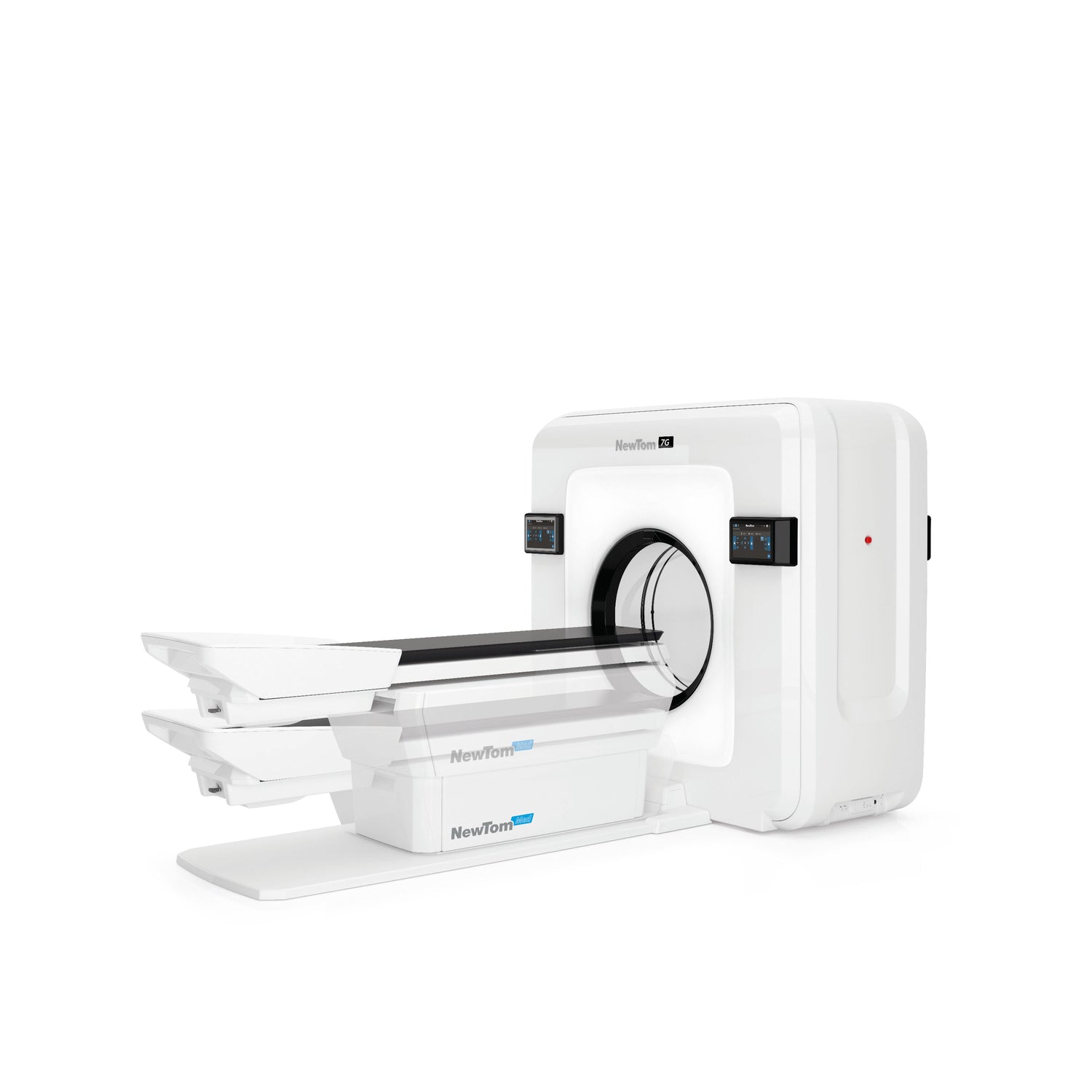
How do we define supine CBCT? Supine refers to a lying down position where the patient is lying on their back while taking an image. CBCT uses cone-shaped X-ray beams and a detector to produce 3D images of a specific part of the body being scanned. Supine CBCT, therefore, refers to a CBCT scan performed while the patient is lying flat on their back.

So, why does supine CBCT produce the best image quality on bone tissues, including micro-structures?
-
Reduced motion artifacts: when a patient is standing or sitting during imaging, there is a higher chance of motion artifacts, which can result in blurred images. When a patient is lying supine, she is less likely to move, reducing the risk of motion artifacts and so producing clearer images.
-
Better control of head position: The supine position allows for better control of the position of a patient's head, which is essential for imaging the maxillofacial region. This can result in better image quality and thus more accurate diagnosis and treatment planning.

-
Reduced scatter radiation: There is a higher chance of scatter radiation when a patient is standing, and this can reduce image quality. In the supine position, the distance between the patient and the detector is greater thereby reducing the amount of scatter radiation and producing clearer images.
-
Consistency in imaging: The supine position allows for more consistency in imaging, as the patient is in the same position for each scan. This can be particularly important for monitoring changes over time, such as in orthodontic treatment.
- Increased patient comfort: And lastly, the supine position can be more comfortable for the patient, reducing the risk of movement during imaging and improving the overall experience.
In conclusion, supine CBCT produces the best quality image on bone tissues, including micro-structures because it reduces motion artifacts, better controls the positioning of the head, reduces scatter radiation, captures consistent images, and allows for greater patient comfort. It is important for clinicians to consider the patient’s position during imaging to ensure the highest quality images are captured to provide the most accurate diagnosis and effective planning of treatment.