


NewTom
NEWTOM 7G
- FEATURES
- SPECIFICATIONS
- DOWNLOADS
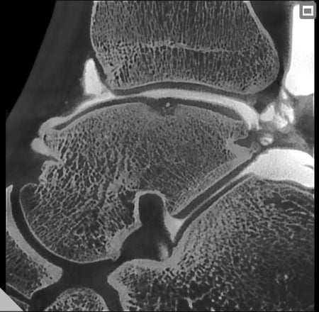
- CLINICAL IMAGES
The first multi-scan body Cone Beam CT now with Dual Energy technology.
The 7G Dual Energy is the first CBCT system capable of imaging the entire body, including the head, spine, shoulders, and hips. This next-generation system can be used to investigate innumerable anatomical areas in a wide range of clinical applications, both 3D with numerous FOVs and 2D (also sequentially). The 7G CT with Cone Beam technology lets users create ultra-high definition images to diagnose the micro-structures of the ear or hairline fractures in complex joints. It can also be used to assess post-surgery outcomes (with minimisation of artifacts caused by bone-articular prosthesis or other osteosynthetic devices such as pins.
Dual Energy
Dual Energy tomography employs two distinct radiant energies to acquire two sets of images of the same anatomical area. Soft tissues have different sensitivities to different energy levels which is why the 7G can now deliver advanced image reconstruction for enhanced diagnostic clarity. Virtual Monochromatic Imaging (VMI) reduces beam-hardening and metal artifacts while improving soft tissue visualisation and supporting accurate tissue characterisation. Colour-coded imaging enables semi-automatic tissue segmentation and material differentiation, making potential pathologies easier to identify. In addition, blended imaging combines high and low energy datasets to produce images with both exceptional contrast resolution and minimal noise, ensuring superior diagnostic confidence in every scan.
Optimal Lying Down Position
With the patient lying down, the motor-powered table limits the risk of artifacts caused by uncontrolled movement. It also ensures simple and accurate alignment of FOVs. Furthermore, the 7G stands out due to its extremely low X-ray doses, which are always in proportion to patient build and clinical requirements.
Large Gantry
The large gantry opening increases the scope for diagnosis and is suitable for heavy patients up to 215kg. Openings on both sides prevent patients from experiencing claustrophobia, while access from the rear also allows use by wheelchair patients.
A FOV for Every Need
The 7G has 15 FOVs, extendable with eXtra Functions. Each FOV is associated with 4 protocols: Low Dose, Regular, Enhanced and Best Quality, ensuring X-ray doses are proportionate to actual needs. The eXtra FOV function lets users perform bilateral examination of the hips, spine and lumbar region - at high resolution.
Low X-ray Dose Protocols
For surgical follow-ups and paediatric imaging, the 7G offers adaptive FOVs, Ultra Rapid and ECO Low Dose modes to significantly reduce X-ray exposure. Its pulsed emission CBCT technology activates radiation only when needed, while SafeBeam™ technology automatically adjusts output to the patient’s anatomy, preventing overexposure. These intelligent dose-control systems enable comprehensive volumetric imaging with radiation levels comparable to just two traditional X-rays. Radiologists can further fine-tune emission settings or begin with a low-dose Ray2D scan, followed by a targeted high-resolution 3D examination for detailed assessment.
Specialist Software
A revolutionary interface makes image display easier and allows for formulation of an immediate diagnosis. Innovative 3D and 2D analysis functions allow pathologies to be identified quickly and accurately, thus optimising workflows whatever the field of application.