One of the most valuable technologies available in dentistry today is the dental operating microscope (DOM). Although many general dentists have historically viewed it as a necessity only for endodontic procedures, the DOM is now gaining wider acceptance. Universities have begun incorporating DOMs into programs outside endodontics, and it is becoming more common for undergraduate programs to feature access to DOMs. They provide numerous benefits to doctors, including the flexibility to adjust focal distance without contorting the body. This allows the doctor to remain comfortable in a more ergonomic position. The DOM dramatically increases visibility, with multiple levels of magnification at the turn of a dial. The enhanced visibility offered by the DOM allows shadow-free lighting. Finally, the DOM facilitates communication between the doctor and patient, as well as the doctor and assistant, through digital documentation and imaging capabilities.
Advantages and Benefits of Dental Operating Microscopes
1. Increased Visualization = Magnification + Illumination
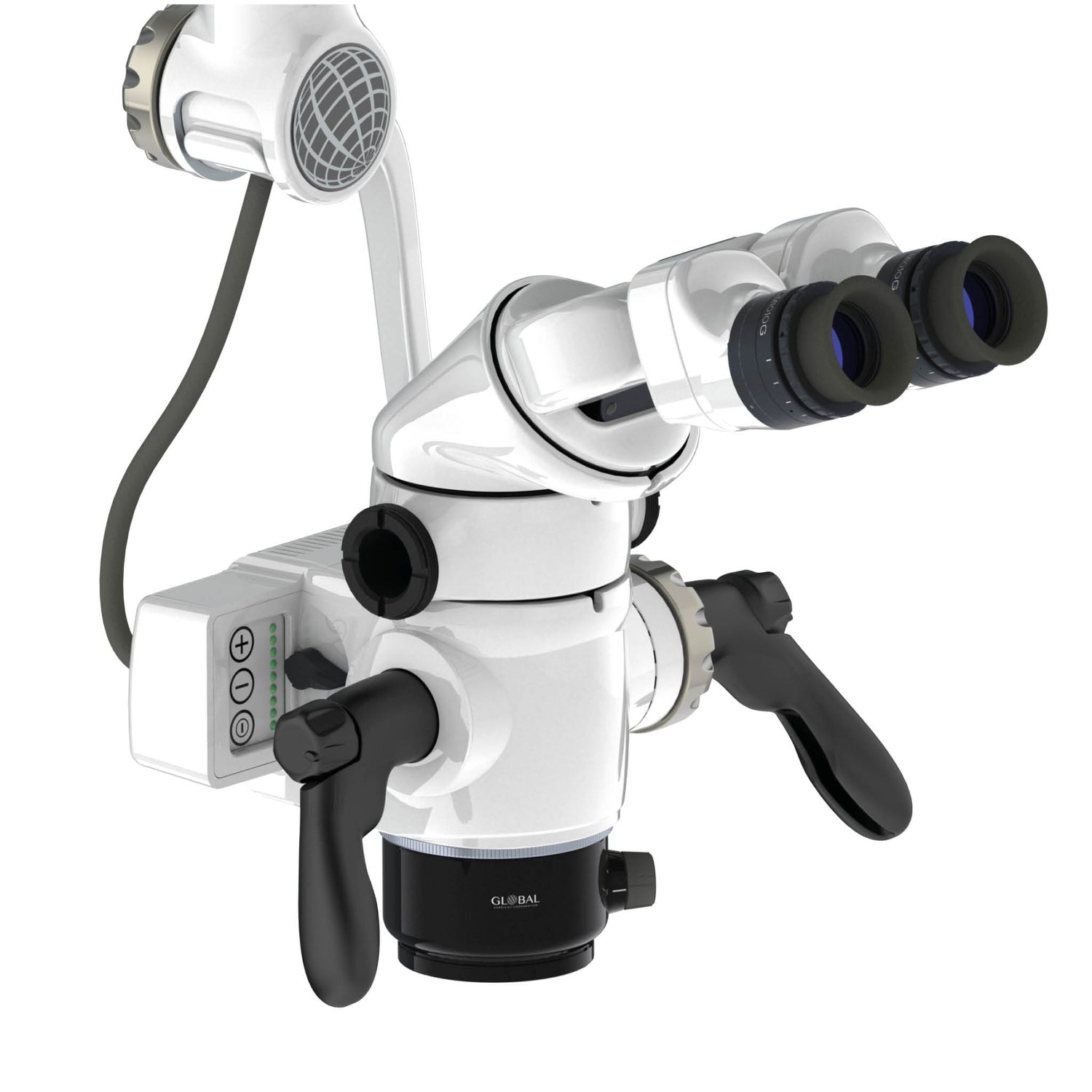
DOMs provide up to six levels of magnification, ranging from 2x to 20x. Another critical component in increasing visualization is illumination. Most microscopes are equipped with an integrated coaxial light source that allows unobstructed, shadow-free illumination of the operating field. With coaxial illumination, the path of light is directed parallel to the DOM's optical axis, which allows significantly improved visualization of even the most difficult-to-access areas of the oral cavity.

With enhanced visualization, the clinician's ability to diagnose pathology in the earlier stages of a disease process is possible. Treatments can also be performed with greater precision, thereby reducing the occurrence of failures and the need to redo procedures. Enhanced visualisation can also allow treatment to be provided more comfortably to the patient because of the clinician's lighter, more refined hand movements, which occur naturally when the clinician is accustomed to operating in a well-illuminated magnified field. Increased visualisation can ultimately result in greater efficiency and productivity because less time is wasted with tactile exploration and checking that all decay has been removed or that a crown margin is fully seated. When a clinician can see all areas of the mouth or a tooth perfectly, the level of efficiency and precision in diagnosis and treatment naturally increases. Simply stated, one cannot effectively diagnose or treat what one cannot see.
2. Communication
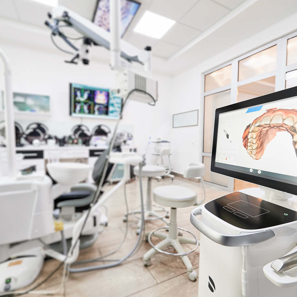
Improved communication is perhaps one of the most notable advantages that DOMs offer in comparison with loupes. A significant return on investment can potentially be realised, provided that the DOM is fully integrated and used to its full potential. Most DOMs offer options for integrating various types of digital recording devices, such as a digital single-lens reflex camera (DSLR) and/or video camera. Digital documentation capabilities enable the clinician to efficiently capture and share with patients what is seen during an examination preoperatively, during treatment intraoperatively, and postoperatively.

Images captured during an examination can be used as a communication and education tool in helping patients better understand their diagnostic findings and why certain treatments may be necessary, especially for conditions that produce no obvious symptoms. Allowing a patient to see the images can lead to greater rates of case acceptance and can significantly streamline the amount of time required in gaining it.
During treatment, images can be efficiently captured, shared, and stored in a patient's digital chart. This is especially useful when unforeseen issues are encountered, such as extensive decay and cracks. Images not only help to increase a patient's trust and confidence in the treating doctor, especially for newer patients, but can also aid in reducing one's medico-legal risk
Additionally, a live video source can be attached to the microscope and fed to a TV or computer monitor, strategically positioned so the dental assistant can easily view it. When the assistant can see exactly what is being performed during a procedure, the assistant's efficiency will increase, and interest and motivation will also rise dramatically because the assistant will tend to feel more involved during the procedure.
After treatment is completed, a clinician may want to provide patients with preoperative, intraoperative, and postoperative photographs of their treatment to emphasise the results. Giving the photographs to patients may have the added benefit of stimulating referrals of patients' friends and family members. The photographs can be given to patients by either printing or emailing them.
In addition to improving communication and education for patients, preoperative photographs taken during an oral examination can also be useful in gaining insurance approval for proposed treatment, minimising or eliminating the need to write narratives. Although photographs can also be obtained with an intraoral camera, the quality of the images and the ease in capturing them is more efficient with a DOM.
3. Improved Ergonomics
Ergonomic improvement is perhaps the most important benefit of the DOM because it allows the clinician to practice with less pain. The prevalence of neck pain among dentists hovers around 70%. Working with a neck posture with only a 20° forward angle has been associated with neck pain. By design, DOMs help to facilitate a near neutral head and neck posture. This is important because most dentists and hygienists operate with a forward head posture of at least 30° for 85% of their time in the operatory.

By operating in a more upright, neutral posture position, the operator is less likely to experience fatigue or strain of neck and back muscles and is, therefore, able to work comfortably for extended periods of time. This in turn can enable the practitioner to provide more dentistry in fewer visits, increasing the clinician's productivity and creating happy patients.
Dental Operating Microscopes Versus Loupes
Dentists have long understood the importance of magnification, as evidenced by the widespread popularity of loupes. There are many obvious differences of the DOM from loupes, such as size, cost, portability, multiple levels of magnification, image stability, and the ability to integrate cameras. Less obvious but significant differences include the following:
- DOM users have very little eye strain and fatigue compared with loupe users, especially as magnification levels increase. This can be attributed to the difference in optical lens design between the two. The DOM relies on infinity-corrected optics that allow stereoscopic vision, whereas loupes rely on convergent optics.
- A DOM offers unobstructed, shadow-free coaxial lighting that is emitted directly from the objective lens parallel to the line of vision. Lighting that is attached to loupes or a headlamp is not coaxial and, therefore, not completely shadow-free.
- A DOM enables dentists to operate in an ideal, fully upright postural position with minimal to no stress or fatigue on the musculoskeletal system, especially the neck and back. Although loupes do help in improving postural ergonomics when compared with unassisted vision, some forward inclination of the head and neck is unavoidable, and the sheer weight of the loupes along with accessory lighting adds additional stressors to the musculoskeletal system.
Click here to LEARN MORE about the Features of the Global Dental Microscope