
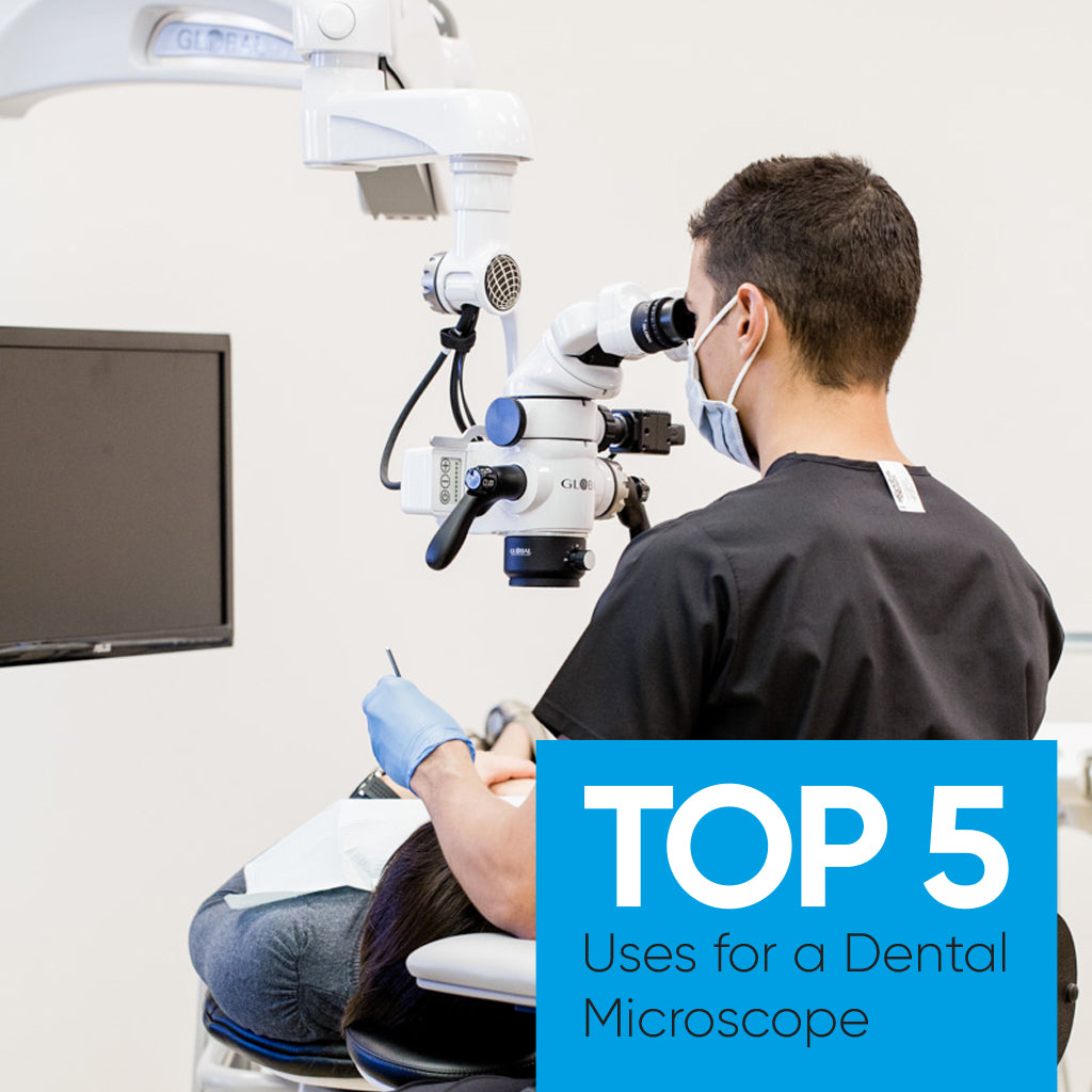
According to Global Surgical, world pioneers in dental microscopy, the following are the top 5 most common uses for a dental microscope and how it benefits your practice.
- General Dentistry
- Endodontics
- Restorative Dentistry
- Documentation
- Patient Education
As services offered by General Dentists expand, the popularity of investing in dental microscopes has increased. Availability of and interest in training in dental specialties outside of basic dentistry, as well as advanced precision and accuracy offered by a microscope, has meant additional services are now being offered to patients, reducing the need to refer out of practice for specialty procedures.
And just like CBCT revolutionised dental X-ray imaging, the dental microscope has done the same for magnification in dentistry. By enhancing visibility with magnification up to 20 times what the naked eye can see, integrated lighting to reduce shadows and photo/video recording attachments, patients can be treated more effectively.
Endodontics
Magnification is accepted as required nowadays to perform endodontic procedures effectively. Root canals can be extremely small and while loupes have been utilised by some, a dental microscope offers significantly improved visualisation and ergonomics.
Improved illumination and magnification enhance the precision and manipulation of all diagnostic and technical aspect of endodontics. In particular:
- Diagnosis: improves visualisation of fracture patterns, both coronal aspect of the tooth, the chamber, and into the root structure.
- Isolation, evaluation, and remediation of pulpal chamber obstructions, calcifications, and resorptions.
- Allowing for predictable pulp chamber visualisation, the investigation of calcified anatomy can be conducted safer, with reduced loss of tooth structure, minimising iatrogenic incidents.
- Three-dimensional visualisation of root obstruction from separated instruments allowing for more successful removal of iatrogenic blockages.
- Endodontic retreatment cases successfully “disassembled” due to predictable removal of core build-ups, cemented posts, silver cones, and previous root fill materials.
- Careful evaluation of biomechanics allowing for more complete cleansing and disinfection of root canal systems.
- Thorough cleansing of deep caries can be completely illuminated, removed, and sealed from oral fluids.
- Manipulation of gingival tissue, for example electrosurgical, suturing, and laser procedures can be more delicately directed with enhanced visualisation.
- Endodontic surgical cases are enhanced as the complete lingual aspect of canal root structure can be more readily seen, allowing for complete root resection, and retro fill procedures. Retro fill preps can be directed to their proper alignment and depth. Enhanced surgical visualisation allows for careful evaluation of suspected fractured root structure and perosseous defects.
Restorative Dentistry
Dental microscopes greatly aid the restorative workflow by providing detailed images of the oral tissue, teeth and gums and thus ensuring more accurate diagnosis. Specifically:
- Provides refinement in tooth and margin preparation.
- Allows for closer inspection of restorations and marginal tissues.
- Improved lighting and magnification aid in caries detection and removal.
- Improves detection and evaluation of coronal and root fractures and abnormalities.
- Facilitates cord placement for gingival retraction.
- Provides for better inspection of impressions.
- Helps with inspection of marginal fit of restoration (crowns, veneers, inlay/on lay, amalgam, composite).
- Facilitates in finishing and polishing of margins.
- Assists in gingival contouring or reshaping around teeth and implants.
- Assists in evaluation after cementation.
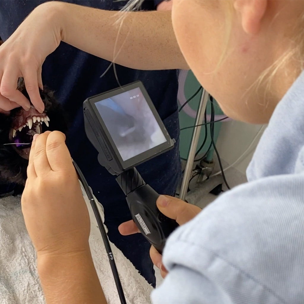
Dental professionals across the globe are using video and photographs in their practices to record treatments, which is made easy with a dental microscope. They give you the ability to record and document procedures on the fly without stopping to switch devices and can integrate with digital single-lens reflex (DSLR) cameras and HD video sources.
In addition, the video/photo functionality is useful for submitting insurance reimbursements, adding video to clinical cases/lectures and patient education.
Patient Education
One of the most effective ways microscopes are used in dentistry is as a patient education tool. Showing your patient in real-time and explaining your proposed treatment plan, ensures more immediate treatment acceptance and uptake, and fosters confidence and rapport for repeat business and referral business, directly boosting your bottom line.
Visualisation through high level of magnification helps ensure you’re capturing all the details when providing a diagnosis. Precision is key to effect diagnosis and planning treatment outcomes. And when effectively communicated to your patients, maximises the benefit of using a dental microscope.
References: GLOBAL A-Series Microscope (inline.com.au) and www.globalsurgical.com